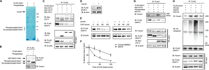
FIGURE 3.
Atoh1 phosphorylation by CK1 is essential for Huwe1-mediated degradation. A and B, HEK cells were transfected with FLAG-Atoh1 for 24 h. Cell lysates were treated with or without λ-phosphatase before immunoprecipitation with a FLAG antibody. Atoh1 and associated proteins were detected by Coomassie Blue staining (A). The areas indicated (40 and 450 kDa) were excised for mass spectrometry and Western blotting (IB) with FLAG (Atoh1) and Huwe1 antibodies (B). IP, immunoprecipitation, C, HEK cells were co-transfected with FLAG-Atoh1 and Myc-CK1 (including CK1α, CK1δ, and CK1ϵ). Immunoprecipitation was performed under non-denaturing conditions with a FLAG antibody. CK1 and Atoh1 were detected with antibodies to Myc and FLAG. Immunoblotting of 5% input is shown below. D, HEK cells were transfected with FLAG-Atoh1 and treated with CK1 inhibitor, D4476 (10 μm), for 18 h. Lysates were processed for Western blotting with FLAG (Atoh1) or β-actin (loading control) antibodies. E, HEK cells were transfected with FLAG-Atoh1 for 40 h and treated with either vehicle (DMSO) or D4476 (10 μm) for 18 h followed by cycloheximide (100 μg/ml) incubation for the indicated times. β-Actin served as a loading control for input protein. F, the ratio from E of Atoh1 to β-actin based on densitometry was plotted. Error bars indicate S.E. Data from three experiments are shown. G, HEK cells were co-transfected with HA-ubiquitin, FLAG-Atoh1, and/or Myc-CK1∂ for 48 h and treated with MG132 (10 μm) for 4 h and λ-phosphatase before immunoprecipitation with a FLAG antibody. Endogenous Huwe1 was detected with an antibody to Huwe1, and Atoh1 was detected with anti-FLAG. Immunoblotting of 5% input is shown below. H, HEK cells were co-transfected with HA-ubiquitin and FLAG-Atoh1 for 48 h and treated with CK1 inhibitors (D4476) and/or proteasome inhibitor MG132 (10 μm) for 4 h. Immunoprecipitation was performed with anti-FLAG antibody. Endogenous Huwe1 was detected with an antibody to Huwe1. Atoh1 was detected with anti-FLAG, and ubiquitin was detected with an antibody to HA. Immunoblotting of 5% input is shown below.

