FIGURE 7.
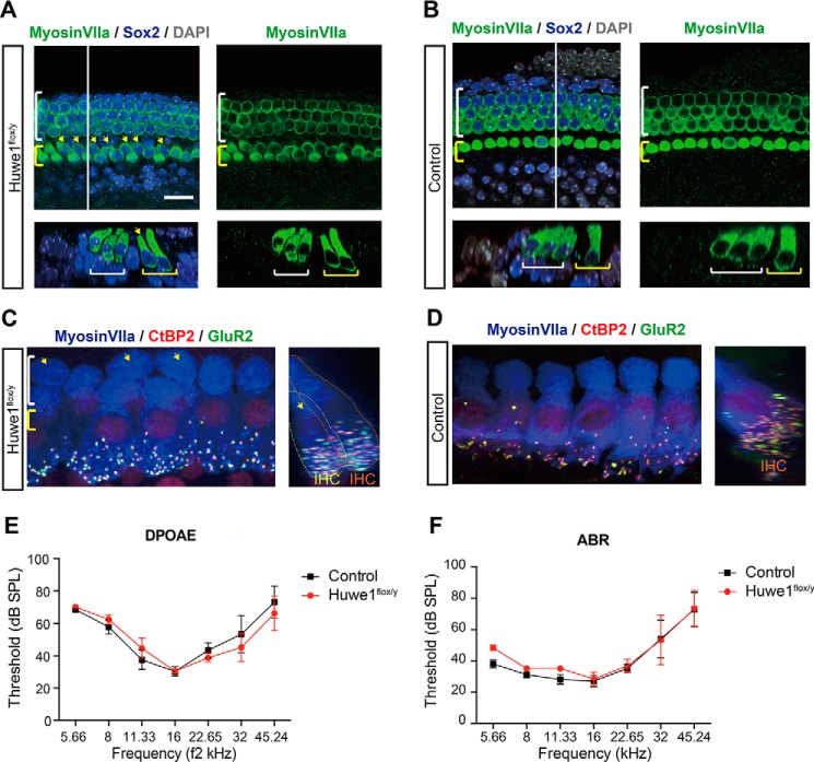
Huwe1 conditional knock-out gives rise to an extra row of inner hair cells. A, yellow arrows indicate additional inner hair cells in the organ of Corti at E19 after knock-out of Huwe1 in Sox2-positive supporting cells (Huwe1flox/y) at E16. Myosin VIIa labels hair cells and Sox2 labels supporting cells; DAPI is a nuclear marker. The white line marks the location of the orthogonal view shown beneath the surface view, and yellow and white brackets indicate inner and outer hair cells, respectively. The scale bar is 25 μm. B, E19 organ of Corti from Sox2-Cre control mice contains a single row of inner hair cells. C and D, Z (left) and X (right) projection confocal images of the inner hair cell region in the P30 organ of Corti from a Huwe1 conditional knock-out (C) or control (D) ear is shown after deletion of Huwe1 at P1. The cochleae were immunostained with antibodies against myosin VIIa, CtBP2, and GluR2. Arrows indicate extra inner hair cells. Yellow and orange dotted lines delineate extra inner hair cells (IHC) on the pillar or modiolar side of the organ, respectively (C, right). E and F, DPOAE (E) and ABR (F) thresholds recorded at P30 from Huwe1 conditional knock-out (n = 3) and control (n = 5) ears are shown. Error bars indicate S.E. SPL, sound pressure level.

