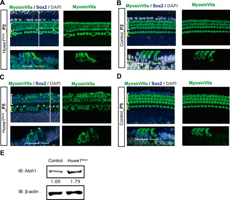
FIGURE 8.
Huwe1 conditional knock-out causes hair cell death. A, enlarged inner hair cells can be seen throughout the cochlea at P2 after knock-out of Huwe1 in Atoh1-positive hair cells. The white line marks the location of the orthogonal view shown beneath the surface view, and yellow and white brackets indicate inner and outer hair cells, respectively. Arrows point to enlarged inner hair cells. Myosin VIIa labels hair cells and Sox2 labels supporting cells; DAPI is a nuclear marker. B, P2 organ of Corti from Atoh1-Cre control mice. C, inner hair cells (arrows) are lost, and outer hair cells (asterisks) are enlarged in a P5 organ of Corti from an Atoh1-Cre;Huwe1flox/y mouse. D, P5 organ of Corti from Atoh1-Cre mice. E, organ of Corti from P5 Atoh1-Cre and Atoh1-Cre-Huwe1flox/y. Atoh1 and β-actin (loading control) were quantified by Western blotting (IB; average Atoh1 levels based on densitometry from two experiments are shown below the blot).

