FIGURE 4.

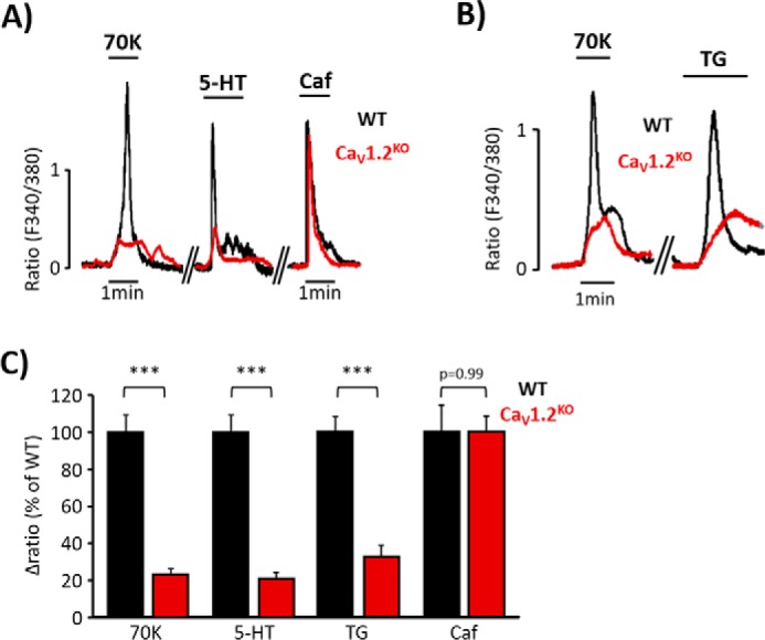
[Ca2+]i increases elicited by serotonin or thapsigargin are diminished in CaV1.2KO mice. A, representative [Ca2+]i changes in aortic VSMC from wild type (WT, black traces) and CaV1.2 knock-out mice (CaV1.2KO, red traces) mice evoked by high KCl (70 mm; 70K), 5-HT (10 μm), and Caf (10 mm). B, representative [Ca2+]i changes of aortic VSMC from WT (black traces) and CaV1.2KO (red traces) mice evoked by 70K and TG (10 μm). C, data summary of [Ca2+]i changes induced by 70K, 5-HT, TG, and Caf in VSMC from WT (black bars; 70K, 100% ± 8.77; 5-HT, 100% ± 13.18; TG, 100% ± 8.57; Caf, 100% ± 14.23; n = 379–1157) and CaV1.2KO mice (red bars; 70K, 22.92% ± 2.82; 5-HT, 20.76% ± 3.94; TG, 26.83% ± 6.18; Caf, 99.88% ± 8.60; n = 190–640). Values are the percentage of mean ± S.E. ***, p < 0.001 versus control.
