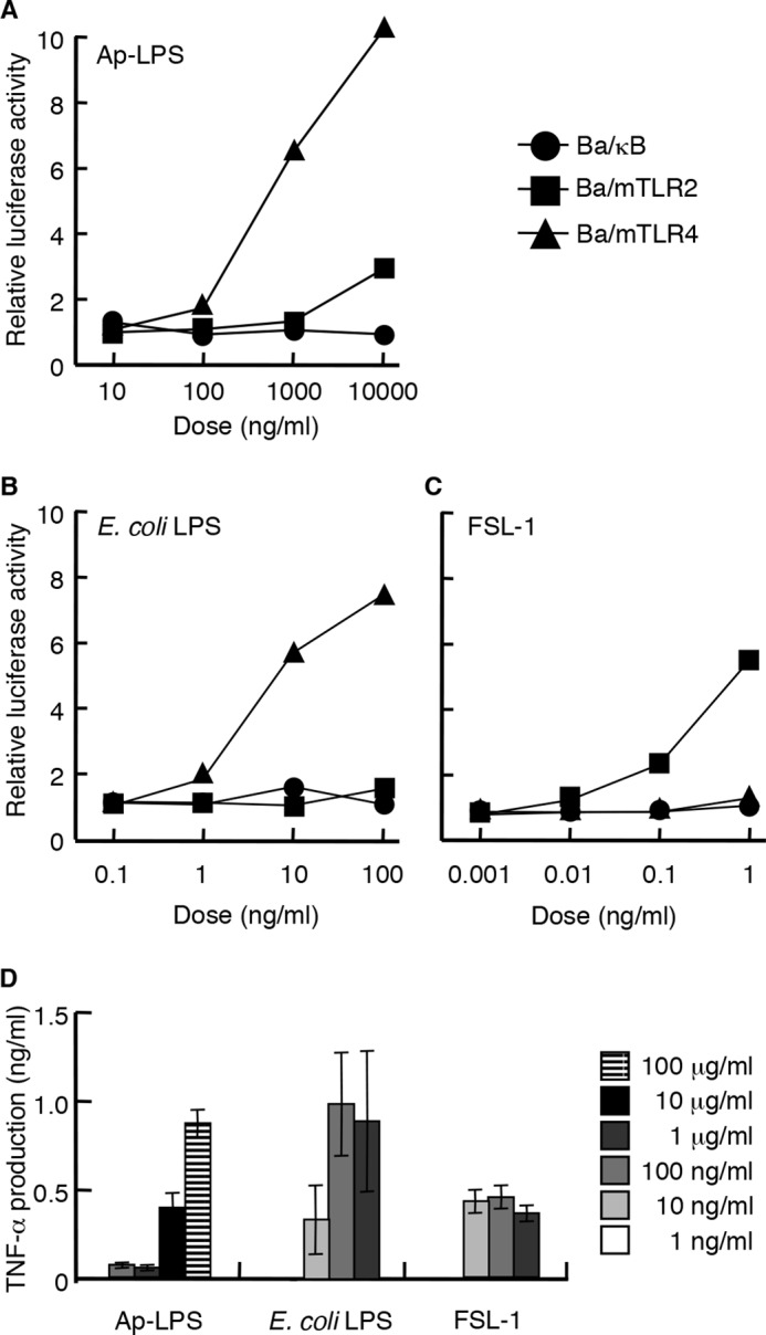
FIGURE 2.

Immunostimulatory activity of Ap-LPS. A–C, the NF-κB activation induced in Ba/κB, Ba/mTLR2, or Ba/mTLR4/mMD-2 cells by Ap-LPS (A), E. coli LPS (B), or FSL-1 (C). The cells were incubated for 4 h, and the activity induced by each reagent was measured with a luciferase assay. D, TNF-α production induced by the indicated concentrations of each stimulus in murine spleen cells. The levels of TNF-α in the culture supernatants of the cells incubated for 4 h were measured by ELISA. E. coli LPS and FSL-1 were utilized as positive controls for TLR4 and TLR2, respectively.
