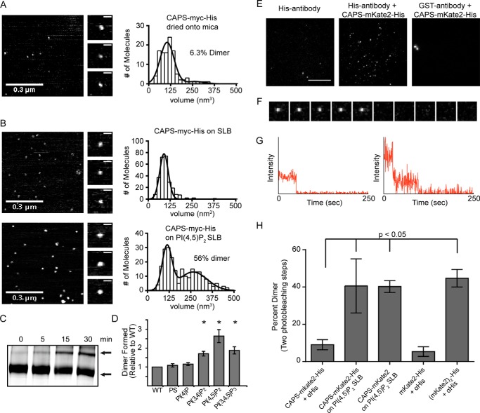
FIGURE 3.
CAPS oligomer is a dimer. A, atomic force microscopy volume analysis of purified CAPS dried onto mica (n = 108). Representative images of AFM field (left), representative images of single CAPS-Myc-His molecules (middle), and histograms of the CAPS-Myc-His volume distribution (right) are shown. Volume data were fit to a double Gaussian with volumes of 109 nm3 for monomer and 264 nm3 for dimer. B, atomic force microscopy analysis of purified CAPS-Myc-His added to SLB (top, n = 448) or SLB containing PI(4,5)P2 (bottom, n = 238) detected monomers at 94 nm3 or monomers at 115 nm3 plus dimer at 261 nm3, respectively. Inclusion of PI(4,5)P2 in SLBs significantly increased the amount of CAPS-Myc-His dimer (p < 0.0001, Mann-Whitney-Wilcox test). C, CAPS-Myc-His dimers increased upon PI(4,5)P2 addition as indicated by BN-PAGE of 5 μg of CAPS-Myc-His incubated with 1.5 μg of PI(4,5)P2 for 0–30 min. Arrows, high mobility monomer and low mobility dimer. Results shown are representative of six experiments. D, incubations and analysis similar to C were conducted with the indicated phospholipids showing that CAPS-Myc-His dimers were preferentially induced (-fold increase is shown) by PI(4,5)P2 (mean ± S.D. (error bars); *, p < 0.001, n = 5). E, pull-down of single CAPS-mKate2-His molecules. TIRF microscopy images showed puncta when CAPS-mKate2-His was retained by His antibody (middle) but not with control antibody (right). Scale bar, 10 μm. F, TIRF images (collected at 2 Hz) showing a one-step photobleach event for immobilized CAPS-mKate2-His. Image size is 2.5 × 2.5 μm. G, representative fluorescence intensity versus time analysis for 1-step and 2-step photobleach events for CAPS-mKate2-His. H, percentages of puncta exhibiting two photobleaching steps were quantified for CAPS-mKate2-His tethered by His antibodies (n = 486), CAPS-mKate2-His bound to PI(4,5)P2 SLBs (n = 296), CAPS-mKate2 (lacking His tag) bound to PI(4,5)P2 SLBs (n = 209), and mKate2-His (n = 307) or (mKate2)2-His (n = 117) tethered by His antibodies. Note that the (mKate2)2-His dimer was only 50% pure and contaminated with fluorescent monomeric mKate2. Values are mean ± S.D.

