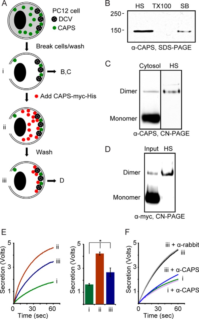
FIGURE 4.

CAPS dimer is the functional form of CAPS. A, PC12 cells were passed through a ball homogenizer (10-μm clearance) to tear the plasma membrane and were washed in KGlu/BSA to remove cytosol (i). Purified CAPS-Myc-His was added to 20 nm (ii), and cell ghosts were washed with KGlu/BSA to remove unbound CAPS-Myc-His (iii). These three states were assessed for activity in E. B, washed cell ghosts (i) were sequentially extracted with 300 mm NaCl (high salt (HS)), 1% Triton X-100 (TX100), and SDS sample buffer (SB) with boiling. Samples were analyzed by Western blotting from SDS-PAGE with CAPS antibody. Results shown are representative of three studies. C, cytosol from PC12 cells (left lane) was compared with HS extract of endogenous CAPS from cell ghosts (i, right lane) in electrophoresis by CN-PAGE and Western blotting with CAPS antibody. Results shown are representative of four studies. D, purified CAPS-Myc-His (1% of input shown) was compared with 50% of HS extract from cell ghosts (iii) in electrophoresis by CN-PAGE and Western blotting with Myc antibody. Results shown are representative of two studies. Endogenous CAPS (C) or exogenous CAPS-Myc-His (D) retained by PC12 cell ghosts migrated as dimer. E, Ca2+-dependent norepinephrine secretion was monitored by rotating disk electrode voltammetry from cell ghosts indicated as i, ii, and iii in A. Ca2+ was injected at zero time. The results from three studies were averaged in the histogram (means ± S.E. (error bars)); *, p < 0.05. F, neutralizing CAPS antibodies inhibited the stimulation of secretion by prebound CAPS (iii). Rabbit IgGs (α-rabbit) were used as the control for CAPS antibodies (α-CAPS). Results shown are representative of two studies.
