FIGURE 7.

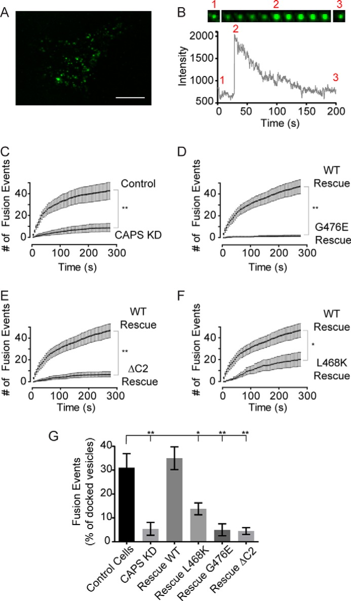
C2 domain mutants exhibit reduced activity in dense core vesicle exocytosis. A, representative TIRF image of PC12 cell expressing BDNF-GFP. Scale bar, 10 μm. B, fluorescence intensity changes for a single vesicle fusion event. Vesicle fluorescence is evident in the TIRF field before fusion (1); fluorescence increases at the time of fusion pore opening (2); and fluorescence subsequently decreases due to reacidification of the cavicaptured vesicle (3). The width of each image in the montage is 1.5 μm. C, exocytosis was evoked by depolarization with the addition of 56 mm KCl buffer at zero time. Curves indicate cumulative number of fusion events per cell. CAPS knockdown significantly impaired secretion compared with control cells (mean ± S.E. (error bars), n = 14). D, rescue studies in CAPS knockdown cells for CAPS-TagRFP (n = 14) or CAPS(G476E)-TagRFP (n = 11). E, similar to D for rescue studies with CAPS-TagRFP (n = 14) or CAPS(ΔC2)-TagRFP (n = 15). F, similar to D for rescue studies with CAPS-TagRFP (n = 14) or CAPS(L468K)-TagRFP (n = 15). The similar expression of CAPS proteins was confirmed by fluorescence of CAPS-TagRFP constructs. Significant differences are indicated: **, p < 0.001; *, p < 0.05. G, summary of CAPS C2 domain mutants compared with wild-type CAPS. Fusion events per cell were normalized to the number of vesicles visible in the TIRF field (mean ± S.E.; *, p < 0.05; **, p < 0.001).
