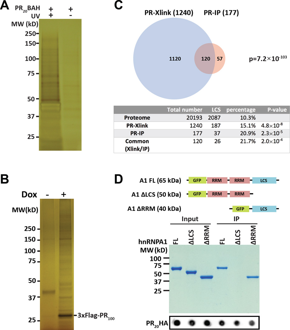
Figure 1. Intracellular targets of the PRn poly-dipeptide.
(A) Silver-stained SDS-PAGE gel of HeLa cell proteins crosslinked to PR20BAH (Experimental Procedures). Left lane shows proteins from cells exposed to both PR20BAH probe and UV light, right lane shows proteins from cells exposed to PR20BAH probe but not UV illumination. Proteins were extracted from the gel and identified by shotgun mass spectrometry (See also Figure S1, Table S1).
(B) Silver-stained SDS-PAGE gel of immunoprecipitated proteins bound by the 3xFlag:PR100 protein conditionally expressed in U2OS cells. 3xFlag:PR100 expression was either induced with doxycycline (+) or not (−). Proteins were extracted from the gel and identified by mass spectrometry (See also Table S2).
(C) Venn diagram showing overlap of 120 proteins commonly identified both by photo-crosslinking to the PR20-BAH probe (PR-Xlink) and immunoprecipitation with the conditionally inducible 3xFlag:PR100 protein (PR-IP). Table below Venn diagram lists the number of proteins associated with low complexity sequences (LCS) in the entire human proteome as compared with the lists of proteins identified to interact with either or both of the PRn probes (See also Table S3).
(D) Coomassie stained SDS-PAGE gel of GFP:hnRNPA1 fusion proteins co-precipitated with the PR20-HA probe. Input and immunoprecipitated PR20HA signals are shown below commassie stained gel as dot blots.

