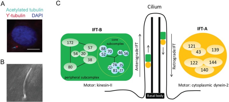
Figure 1.
Primary cilium structure and intraflagellar transport proteins. (A) Immunofluorescence micrograph of primary cilium located on primary osteoblasts derived from mouse calvarial bone. Primary cilium was stained with γ-tubulin (basal body; red) and acetylated tubulin (axoneme; cyan) antibody. Nuclear was stained with DAPI (blue). Scale bars represent 10 μm. (B) Scanning electron microscopic image of primary cilium present on mouse mesenchymal stem cell. Scale bars represent 2 μm. (C) Schema of primary cilium structure and intraflagellar transport (IFT) complexes. Adapted from Katoh et al. (2016). This figure is available in color online at http://jdr.sagepub.com.

