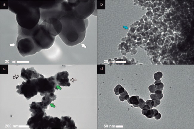
Figure 2.
Transmission electron microscopy images of the nanopowders: (a) Silica-coated zirconia particles (×1.2M magnification). The white arrows point to zirconia nanoparticles (darker area at the center) surrounded by a silica layer (grayish area). The thickness of the silica layer seems to vary according to the diameter of the zirconia particles, which is visible when the 2 examples indicated by the white arrows are compared. (b) Silica-coated alumina particles (×1M magnification). Discrete nanoparticles much smaller than the other particles are observed. The blue arrow points to an example of a coated particle. (c) Silica-coated titania particles (×100k magnification). Most particles are seen embedded into clusters with a silica matrix (hollow black arrows), though some coated particles are also present (green arrows). (d) Silica nanoparticles (×300k magnification) were detected, most likely as a result of a secondary phase formation during heat treatment of tetraethylorthosilicate molecules that were not bound to the nanopowders in the solution used for the silica-coating method. This figure is available in color online at http://jdr.sagepub.com.

