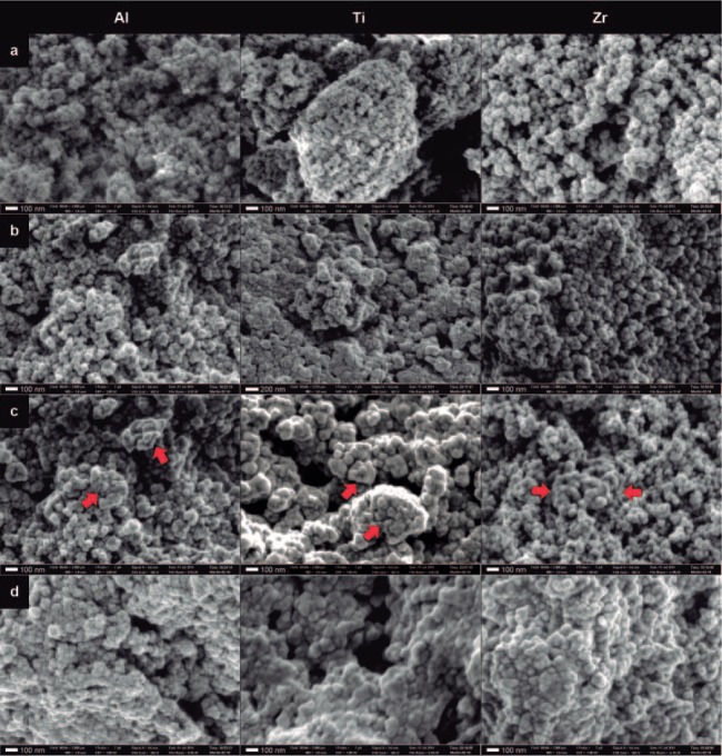
Figure 3.
Field emission scanning electron microscopy images of the nanopowders. Imaging conditions: electron beam set at 1 kV and 7 pA, sample distance 3 mm, and imaged in a field width of 2 μm—equivalent to approximately ×180k magnification: (a) Image of the nanopowders as received. Alumina (Al) particles are visibly smaller than the other particles. (b) Image of the nanopowders after silica coating. Most of the titania (Ti) particles are embedded into clusters with a silica matrix, which does not seem to happen with the other particles. All particles became highly unstable after silica coating (c), where some areas in the field (red arrows) show the particles fused as a reaction to the energy of the microscope beam. This is clearly seen for the silica-coated zirconia image, where the fusing started near the reduced focus area in the center of the field. (d) The uncontrolled fusing of particles, caused by the energy of the beam and taking over the entire field, thereby generating a 3-dimensional porous structure. This figure is available in color online at http://jdr.sagepub.com.

