Figure 2.
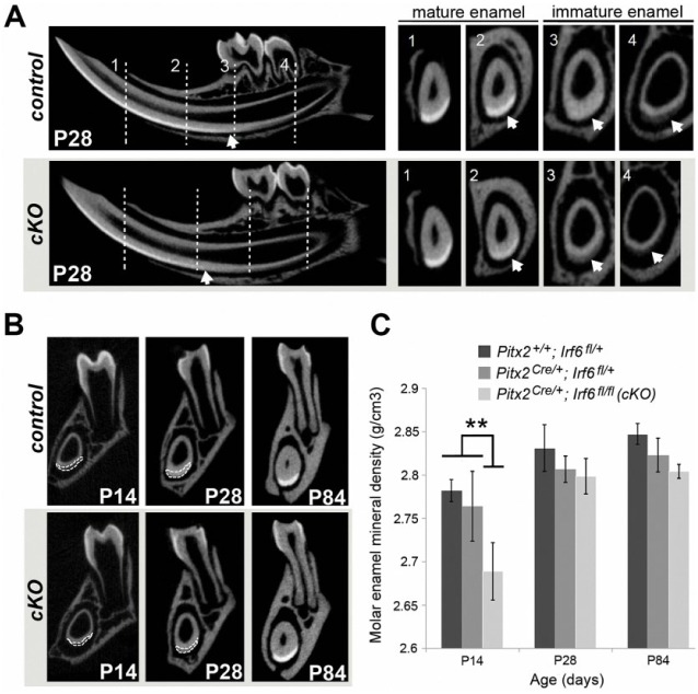
Delayed enamel maturation in Irf6-cKO (conditional knockout of Irf6) mice. (A) Microcomputed tomography imaging confirmed delayed enamel maturation in postnatal day 28 (P28) Irf6-cKO incisors as compared with controls. Representative cross-sections of incisors show 4 landmark regions (vertical lines), including immature enamel at the distal root of the second molar (4), mesial root of the first molar (3), mental foramen (2), and mature enamel at the alveolar crest (1). The transition between immature and mature enamel is incisally displaced (i.e., delayed) in Irf6-cKO animals, as evident when enamel radiolucency is compared in regions 3 and 4 and, to a lesser degree, in region 2 (see arrows). No significant differences were detected between control genotypes. (B) Microcomputed tomography imaging reveals that Irf6-cKO mice exhibit thinner mandibular incisor enamel (outlined in white) versus controls at P14, although incisor enamel radiolucency was comparable by P84 (data not shown). (C) Quantitative analysis of molar enamel mineral density confirmed a significantly decreased (asterisk; P < 0.05) enamel mineral density in Irf6-cKO mice versus controls at P14. However, by posteruption stages (P28 or P84), the enamel mineral density in Irf6-cKO mice was no longer significantly different from that of the Pitx2-Cre heterozygote controls, although it was still significantly lower than in wild-type mice.

