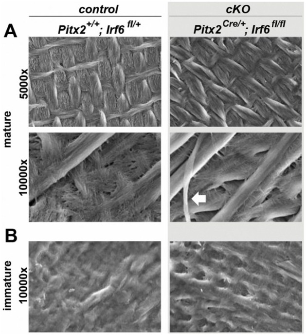
Figure 3.

Scanning electron microscopy analysis reveals a normal enamel prismatic structure in Irf6-cKO (conditional knockout of Irf6) mice. Scanning electron microscopy analysis of postnatal day 28 mice reveals a comparable prismatic structure in control and Irf6-cKO mice in regions of mature enamel (A), as well as a similar appearance of immature enamel (B). However, some shearing of enamel rods was observed in Irf6-cKO samples (white arrow), which was not seen in controls.
