Figure 3.
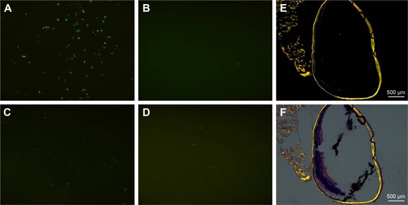
Chemotaxis of the GFP-labeled CR2-sFlt 1 toward complement cleavage fragments in the transwell assay (A–D) and mouse CNV model (E and F).
Notes: (A) GFP signal of CR2-sFlt 1 suspension in the upper chamber before incubation. (B) No GFP signal in the upper chamber suspension after 1 hour incubation. (C) Most increased green signals in the interface membrane of the transwell chamber after 1 hour incubation. (D) Green signals were detected in the lower chamber suspension after 1 hour incubation. (E) Green fluorescence signals in frozen sections of eye tissue in the CR2-sFlt 1-treated group. (F) Green fluorescence signals merged with corresponding HE staining.
Abbreviations: GFP, green fluorescent protein; CNV, choroidal neovascularization; HE, hematoxylin and eosin.

