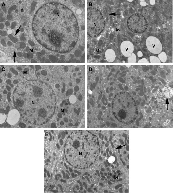
Figure 5.
Electron micrographs of liver sections of rats. (A) Control shows hepatocyte with normal cellular organelles and intact nuclear chromatin; N indicates nucleus, M indicates mitochondria, and arrows indicate endoplasmic reticulum. (B) Arsenic observe increased peripheral chromatin in the nucleus (N), mitochondria (M) with ill-defined cristae, few endoplasmic reticulum (arrow) and numerous obvious vacuoles all over the cytoplasm (V); bc indicates bile canaliculus. (C) Arsenic + NAC: note that less condensed chromatin in the nucleus (N), scattered mitochondria (M), and in-between short strands of endoplasmic reticulum are visible. (D) Arsenic + DMSA: the cytoplasm is heavily populated with mitochondria and less fragmented endoplasmic reticulum (compared to B). Notable increase in organelle degeneration is still evident (arrow). (E) Arsenic + NAC + DMSA: hepatocyte is preserved without vacuolization, nucleus (N) without abnormal chromatin, normal mitochondria (M) with intact cristae, normally shaped rough endoplasmic reticulum (arrow). Original magnification 7,500× (A) and 5,000× (B–E).
Abbreviations: NAC, N-acetylcysteine; DMSA, meso-2,3-dimercaptosuccinic acid.

