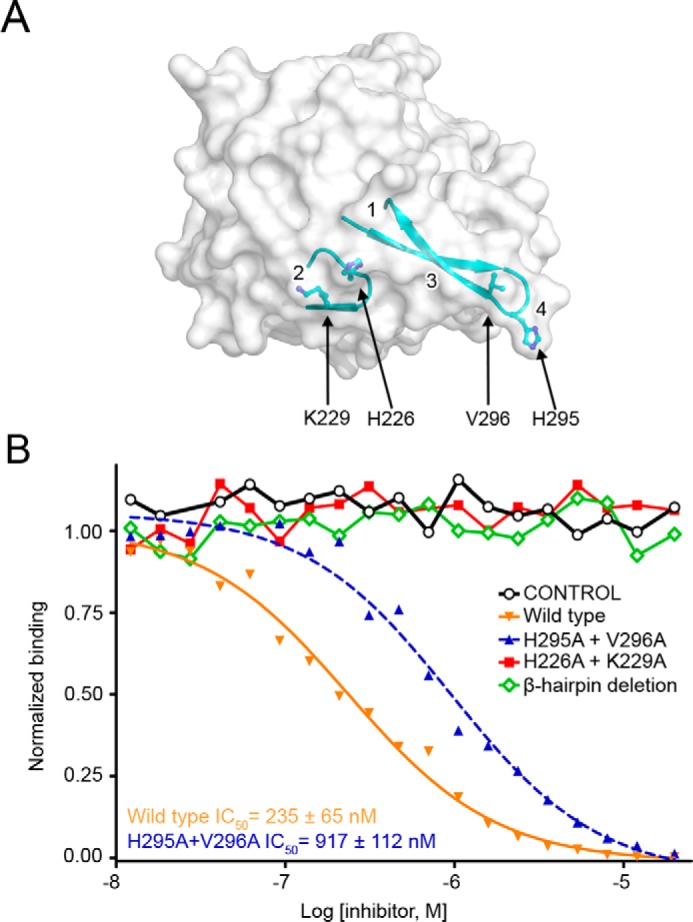
FIGURE 4.

Validation of the PTPRG·CNTN interface. A, surface representation of the CA domain of PTPRG with the two loops responsible for CNTN binding shown in a ribbon representation. The residues mutated to alanine in the binding assays shown in B are shown as ball-and-stick representations. Binding sites 1–4 are labeled on the PTPRG surface. B, mutational analysis of interactions between the CA domain of PTPRG with CNTN4. The ability of bovine CAII (control), mouse PTPRG(CA), or mouse PTPRG(CA) mutants to inhibit binding between an IgG Fc fusion of mouse CNTN4 and a biotin-labeled PTPRG(CA) was assessed over a logarithmic dilution series. See supplemental Fig. S2.
