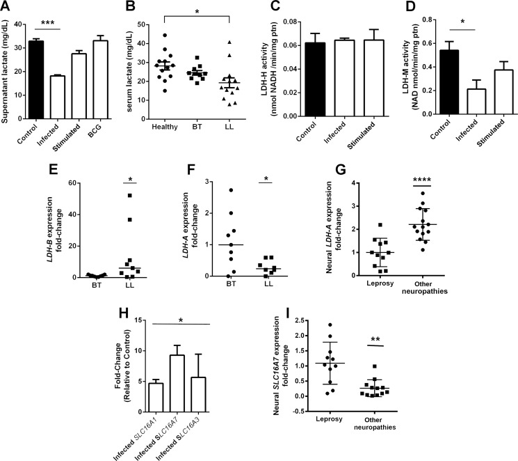
FIGURE 3.
Schwann cell lactate release is affected during M. leprae infection. Lactate release in infected Schwann cell supernatant is reduced by half in comparison to control cells (A). Cells stimulated to BCG or M. leprae inactivated by radiation showed no reduction in lactate. Leprosy patients from the lepromatous pole of the disease (LL) also demonstrated a 30% reduction in serum lactate levels (B). Although Schwann cell lactate dehydrogenase isoform H activity is not affected (C), isoform M activity is reduced during M. leprae infection (D). LDH-B and LDH-A mRNA expression were determined in skin lesions from leprosy tuberculoid (BT) and lepromatous (LL) patients (E and F). LDH-A gene expression was determined in peripheral nerves biopsies from pure neural leprosy patients and patients suffering from other neuropathies (G). As expected, the LDH-A gene is repressed in leprosy patients. The monocarboxylate transporters SLC16A1, 16A3, and 16A7 are up-regulated in infected Schwann cells (H), whereas SLC16A3 is increased in peripheral nerves from leprosy patients in comparison to nerve biopsies from other neuropathies (I). The results are expressed as the mean ± S.E. from three normalized independent biological replicates. Statistical significance was calculated by ANOVA followed by Bonferroni test where: ***, p < 0.0001; **, p < 0.001; and *, p < 0.01 in comparison to control.

