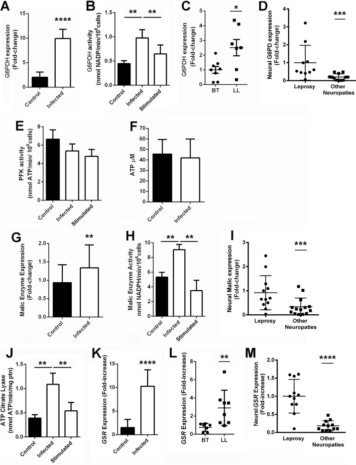
FIGURE 4.
M. leprae induces the Schwann cell pentose pathway oxidative phase. G6PDH activity (A) and expression (B) is increased in infected Schwann cells. G6PDH expression is also increased in lepromatous leprosy skin lesions (LL), where M. leprae is abundant, compared with tuberculoid ones (BT) (C). G6PDH is also increased in peripheral nerves from leprosy patients when compared with nerve biopsies of other neuropathies (D). The PFK (E) and cellular ATP abundance (F) are not altered in infected Schwann cells. On the other hand, malic enzyme, another important source of Schwann cell reducing power, presented increased expression (G) and activity (H) during infection. Malic enzyme is also increased in peripheral nerves from leprosy patients when compared with nerve biopsies from patients diagnosed with other neuropathies not related with leprosy (I). ATP citrate lyase, the first enzyme of the lipid synthesis pathway, is also increased in infected Schwann cells (J). Glutathione reductase, the main destination of cell reducing power, is also increased during M. leprae infection in human Schwann cells (K), skin (L), and nerves (M). Cells incubated with γ-inactivated M. leprae are identified as stimulated. The results are expressed as the mean ± S.E. from three normalized independent biological replicates. Statistical significance was calculated by ANOVA followed by Bonferroni test where: ****, p < 0.0001; ***, p < 0.001; **, p < 0.01; and *, p < 0.05 in comparison to controls.

