FIGURE 3.
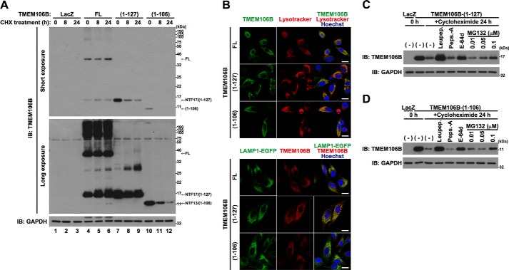
TMEM106B-NTFs are rapidly degraded by lysosomal and proteasomal degradation pathways. A, HeLa cells, seeded on 6-well plates at 5 × 10⁴ cells/well, were infected with adenovirus vectors encoding TMEM106B-FL at an MOI of 800 or TMEM106B-(1–127) or TMEM106B-(1–106) at an MOI of 200. At 18 h after the start of infection, cells were treated with 5 μg/ml cycloheximide (CHX) for 8–24 h. Then the cells were harvested for immunoblotting analysis (IB) using the TMEM106B antibody. B, top, HeLa cells overexpressing non-tagged TMEM106B-FL, TMEM106B(1–127), or TMEM106B(1–106) were incubated with 100 nm Lysotracker (red) at 37 °C for 1 h just before fixation. Then the fixed cells were immunostained with the TMEM106B antibody (green). Bottom, HeLa cells overexpressing LAMP1-EGFP (green) and non-tagged TMEM106B-FL, TMEM106B(1–127), or TMEM106B(1–106) were fixed and immunostained with the TMEM106B antibody (red). Nuclei were stained with Hoechst 33258 (blue). White bar, 20 μm. C and D, HeLa cells, seeded on 6-well plates at 5 × 10⁴ cells/well, were infected with adenovirus vectors encoding TMEM106B(1–127) (C) or TMEM106B(1–106) (D) at an MOI of 200. After the infection, the cells were co-incubated with or without 50 μg/ml leupeptin (Leupep.), 10 μg/ml pepstatin A (Peps.-A), 5 μg/ml E-64d, or 0.01–0.1 μm MG132. At 18 h after the start of infection, the cells were co-incubated with or without 5 μg/ml cycloheximide for 24 h in the presence or absence of 50 μg/ml leupeptin, 10 μg/ml pepstatin A, 5 μg/ml E-64d, or 0.01–0.1 μm MG132. At 24 h after the start of the CHX treatment, the cells were harvested for immunoblotting analysis using the TMEM106B antibody.

