
FIGURE 8.
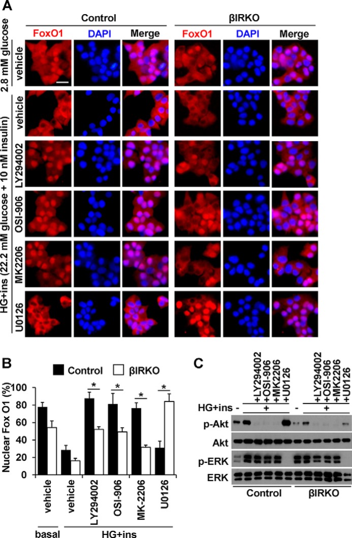
Effects of inhibitors on FoxO1 localization induced by insulin and glucose stimulation in β-cells. After starvation, control or βIRKO β-cell lines were treated with insulin (10 nm) in combination with high glucose (450 mg/dL), in the presence or absence of PI3K inhibitor LY294002, insulin receptor, and IGF1 receptor dual inhibitor OSI-906, Akt inhibitor MK2206, or MEK1/2 inhibitor U0126 for 30 min. A, representative pictures of β-cells immunostained for FoxO1 (red) and DAPI (blue). The scale bar indicates 20 μm. B, proportions of nuclear FoxO1 positive cells in control (filled bars) or βIRKO (white bars) β-cell line. C, Western blotting analysis of indicated proteins in control and βIRKO β-cell lines under the conditions described in A. Data are mean ± S.E. *, p < 0.05 compared with respective controls.