
FIGURE 1.
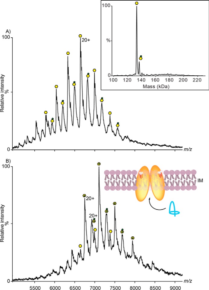
Mass spectrum of McjD. A, mass spectrum of McjD dimer complex exchanged in OGNG detergent acquired on a Q-ToF mass spectrometer. Charge states are assigned to the apo complex (yellow hexagon), phospholipids (yellow hexagon with red adduct), and possibly binding of 3.2–3.5-kDa species of LPS (yellow hexagon with green adduct). In the inset, the deconvoluted mass spectrum of McjD is shown (confirming the mass of the protein). B, McjD incubated with 10 μm lasso-peptide Mccj25 for 10 min. New peaks emerged as adduct to the apo- and LPS-bound McjD showing that binding of lasso peptide is independent of LPS presence. Insets are the position of McjD in the inner membrane and transport of lasso-peptide MccJ25 (shown in blue ribbon) to the periplasmic space. LPS are shown as white spheres and red sticks.