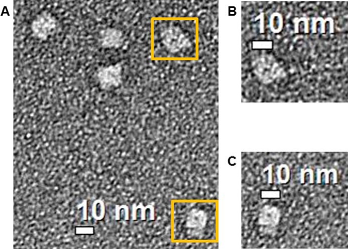
FIGURE 4.

Electron microscopy analysis of the Stt7 tetramer. Diluted Stt7 (2 μg/ml) was absorbed on a carbon-coated 400-mesh copper grid and stained with uranyl acetate. Images were recorded on an FEI Tecnai transmission electron microscope operated at 200 kV at a magnification of ×71,000 and defocus of −1 μm. A, the 2D image shows a square-like organization of the Stt7 tetramer of ∼10 × 10 nm with an ∼14-nm diagonal length. B and C, magnified views of two tetramers of Stt7 highlighted in A with yellow boxes. The tetramers show a dark circular feature at the center that represents a hollow cavity of ∼1 nm diameter.
