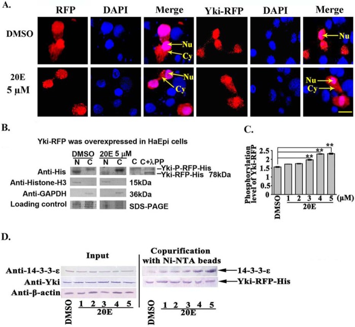
FIGURE 7.
20E arrested Yki in the cytoplasm by regulating Yki phosphorylation and interaction with 14-3-3-ϵ. A, the location of Yki-RFP-His in HaEpi cells. Red fluorescence represents RFP and the recombinant protein annexin-RFP, whereas blue represents DAPI-stained nuclei. Merge shows the superimposed red and blue fluorescence. Nu, nucleus; Cy, cytoplasm. The pictures were obtained after 6 h of 20E (5 μm) incubation; DMSO was used as the solvent control. Pictures were taken using a Zeiss LSM 700 laser confocal microscope. The scale bar represents 20 μm. B, Western blotting analysis of Yki-RFP-His expression in the cytoplasm and nucleus following pIEx-Yki-RFP-His overexpression. The gel concentration was 7.5%. N, nucleus; C, cytoplasm. λPP, λ-phosphatase. Anti-histone-H3 and anti-GAPDH were used to control the separation of the cytoplasmic and nuclear proteins, respectively. Loading control indicated the quantity of the protein loading. C, Yki phosphorylation levels detected using a phosphoprotein phosphate estimation kit. The cells were treated with 20E (1–5 μm) for 6 h. The error bars represent the standard deviations of three replicates. The asterisks denote significant differences (p < 0.01, via Student's t test). D, interaction between Yki and 14-3-3-ϵ (30 kDa). Input, protein expression levels of 14-3-3-ϵ and Yki-RFP-His in the cell lysates after various treatments. The co-purified 14-3-3-ϵ was detected using a rabbit antiserum against Helicoverpa 14-3-3-ϵ prepared in our laboratory. β-actin was used as the quantitative control.

