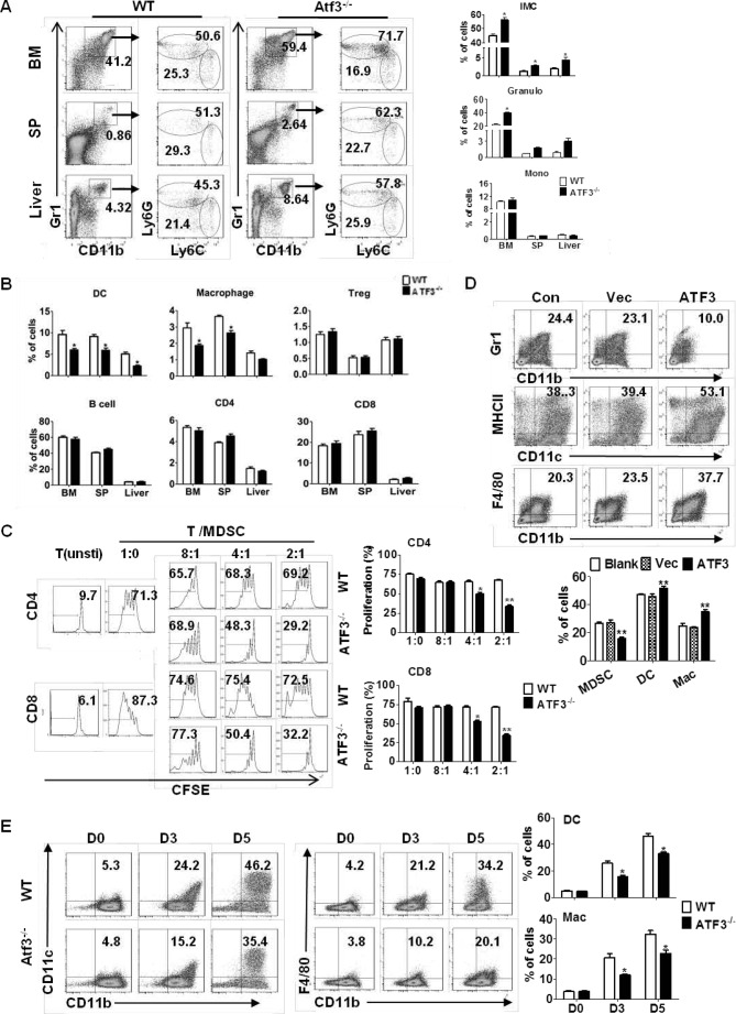
FIGURE 5.
ATF3 regulates the development of G-MDSCs. A, proportions of the indicated myeloid cell populations in WT and ATF3-KO mice were analyzed by flow cytometry. iMC (CD11b+Gr1+), Granulo (granulocytes, CD11b+Ly6G+Ly6C−), and Mono (monocytes, CD11b+Ly6G−Ly6C+) are shown. B, proportions of indicated immune cells in WT and ATF3-KO mice were determined by flow cytometric analysis: DC (CD11c+MHCII+), macrophage (CD11b+Gr1−F4/80+), Treg (CD4+CD25+), B cell (B220+), CD4 T (CD3+CD4+), and CD8 T (CD3+CD8+). Con, control. C, allogeneic mixed lymphocytes reaction (MLR). CD3+T cells were stimulated with ConA and cocultured with allogeneic splenic CD11b+Ly6G+ at different ratios for 3 days. T cell proliferation was evaluated by carboxyfluorescein diacetate, succinimidyl ester (CFSE) dilution, and unstimulated T cells were used as negative control. D, BM cells were infected with lentivirus expressing ATF3 or vector (Vec) and cultured in medium containing GM-CSF and IL6. The frequencies of MDSCs (Gr1+CD11b+), DCs (CD11c+MHCII+), and macrophages (CD11b+F4/80+) among GFP + cells were analyzed by flow cytometry at day 6. Medium alone was used as control. E, splenic MDSCs were cultured with GM-CSF and IL-4 for 3 and 5 days, and the proportions of DCs (CD11c+CD11b+) and macrophages (CD11b+F4/80+) were determined. A–D, both representative data and mean ± S.E. from six mice (A) or three independent experiments (B–D) are shown. *, p < 0.05; **, p < 0.01, unpaired t tests. SP, spleen.

