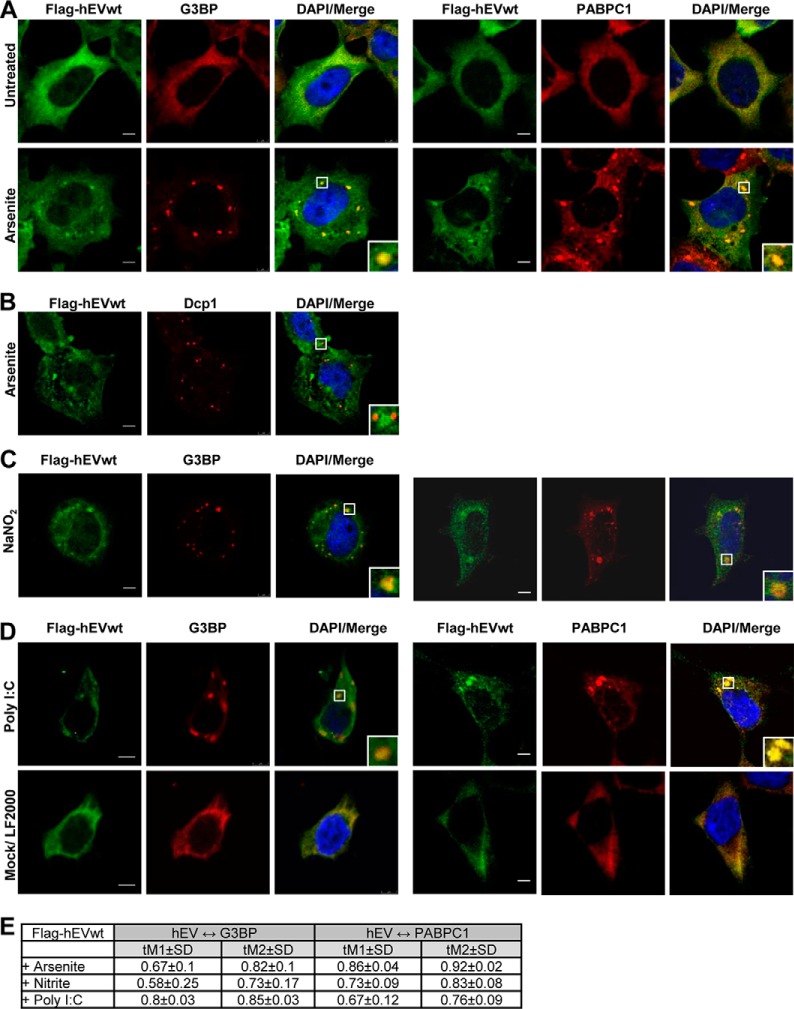
FIGURE 6.
Induction of hEndoV-containing stress granules by various stress. A, T-REx 293 cells ectopically expressing FLAG-hEndoVwt were cultured in the absence or presence of arsenite (0.5 mm, 30 min) before processing for visualization of hEndoV (green) and G3BP (red) (left panels) or hEndoV (green) and PABPC1 (red) (right panels) or (B) hEndoV (green) and Dcp1 (red). C, T-REx 293 FLAG-EndoVwt cells were cultured in the presence of nitrite (200 mm, 1 h) or D, transiently transfected with poly(I:C) (500 ng, 7 h) and processed for imaging of hEndoV (green) and G3BP/PABPC1 (red). Mock-treated cells were also included. Nuclei were counterstained using DAPI (blue) and colocalization (yellow) is shown in Merge. Localization of proteins was observed by confocal microscopy (Leica SP8) using a ×40 oil objective. Bar, 5 μm. E, Manders' coefficients for colocalization of hEndoV and G3BP (left columns) or hEndoV and PABPC1 (right columns).

