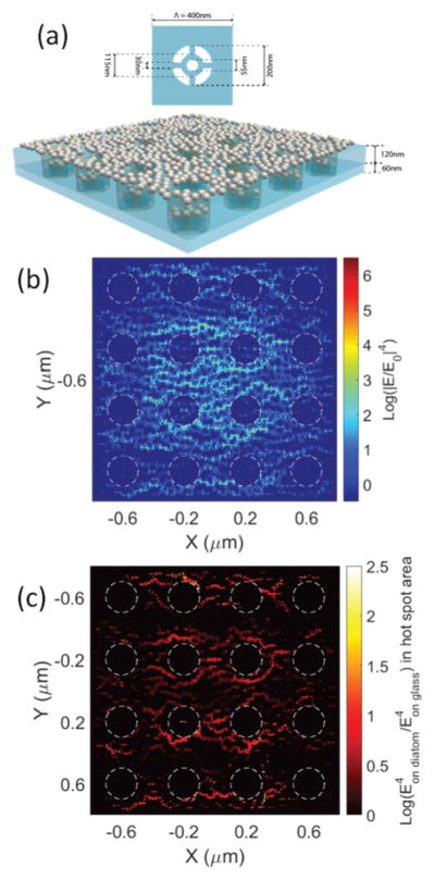
Fig. 1.

(a) Schematic of the 3-D simulated hybrid diatom–Ag NP nanostructures: Ag NPs are randomly distributed on a diatom frustule; (b) 3-D FDTD simulation of the field enhancement |E/E0|4 of the nanostructure; and (c) enhancement factor of the hot-spots compared with those of Ag NPs on a flat glass substrate. Note: the value of |E/E0|4 is plotted in the log-scale.
