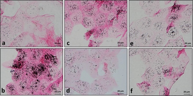
Fig 3. Measurement of melanin remnants in surface corneocytes using Fontana-Masson silver staining.
Surface corneocytes were collected from guinea pig dorsal skin treated as described in Fig 2. Many coarseclustered black silver deposits are seen in corneocytes from vehicle controls (b)and H2O2-treated group (c), whereas fewer silver precipitations are seen in the sham-irradiated control (a) andthe depigmented skin after topical application of 5% HQ (d), 10% arbutin (e) and 10% D-Arb (f) for 10 days. Scale bars: 20 μm.

