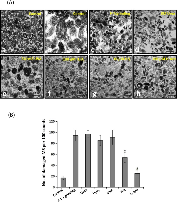
Fig 5. Ultrastructural observations of individual naked melanosomes.
A: Individual naked melanosomes were purified from cultured MNT1 cells, as described under Materials and methods. Typical images of mature melanosomes are shown in low (a) (5000×) and in higher (b) (15000×) magnifications. Melanosome fractions were treated with freeze-thawing (FT) plus manual grinding (c), 8M urea (d), 100 μM H2O2 (e), 3J/cm2 UVA radiation(f), 10 μM HQ (g) and 100 μM D-Arb (h). Significantly fragmented and vacuolated melanosomes are seen in specimens treated with 100 μM H2O2, 3 J/cm2 UVA radiation, and 10 μM HQ (e-g). Scale bar: 1μm (except 0.2 μm for b and 0.5 μm for e). B: Comparison of percentages ofdamaged melanosomes following the different treatments. Two-way ANOVA was used to determine the statistical difference between treated melanosomes and the untreated control. * P<0.05, # P> 0.05.

