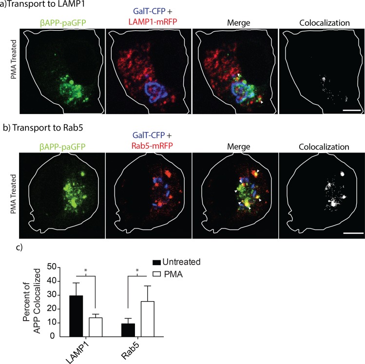
Fig 6. PMA treatment alters the intracellular trafficking of APP.
SN56 cells transiently transfected with βAPP-paGFP were treated or not treated with 300nM PMA for 1-hour before imaging. Cells were photo-activated in the Golgi (GalT-CFP) for 15 minutes. Video of the live cells was taken during this 15-minute period to follow the trafficking of APP. Frames from the beginning and the end of the time course are shown here for transport to a) lysosomes (LAMP1) and b) early endosomes (Rab5). Far-right panels show colocalized pixels between the βAPP-paGFP and LAMP1-mRFP channels. The edge of the cell is defined by the white line, and was drawn based on the white light images. Triangles alone point to colocalized pixels. Scale bars represent 5μm for all images. c) The amount of APP colocalized with each compartment was quantified using Imaris at the 15-minute time point, and the results were plotted using Prism 5.0b. Error bars represent SEM and * denotes p<0.05.

