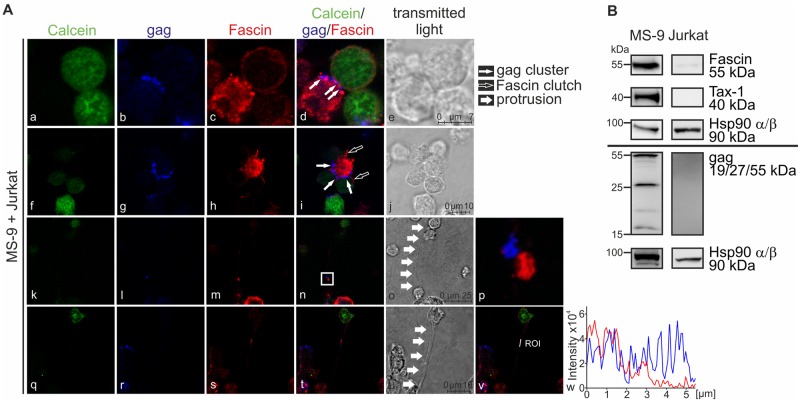
Fig 7. Fascin and gag localize at cell-cell contacts and in long-distance connections between infected and uninfected T-cells.
(A) Confocal laser scanning microscopy of HTLV-1-infected MS-9 cells co-cultured with Jurkat T-cells. Jurkat T-cells were pre-stained with Calcein-AM (green) to differentiate between the two cell types. Cells were co-cultured for 0 (k-p), 30 (a-j) or 60min (q-w) on poly-L-lysine-coated coverslips prior to drying (20min), fixation, and staining. (A) Stainings of Calcein (green), gag (blue), Fascin (red) and the merge of all three stainings are shown. Transmitted light served as control. Representative stainings of three independent experiments showing clusters of Fascin (a-e) and gag (a-j), Fascin clutches (f-j) or long-distance connections (k-w) are depicted. Thin white arrows indicate gag of an infected cell clustering at the cell-cell contact towards an uninfected cell; framed white arrows indicate short-distance Fascin-containing membrane extensions; and thick white arrows indicate long-distance protrusions between uninfected and infected cells. Protrusions (k-o; q-u) were examined in more detail, and (p) the stains of gag and Fascin within the protrusion shown in (n) were enlarged; further, a region of interest (v-w) was analyzed showing the intensities of gag- (blue) and Fascin- specific (red) fluorescences shown in (t). (B) Detection of Fascin, Tax-1 and gag in HTLV-1-infected MS-9 cells and uninfected Jurkat T-cells by western blot. Hsp90 α/β served as control.

