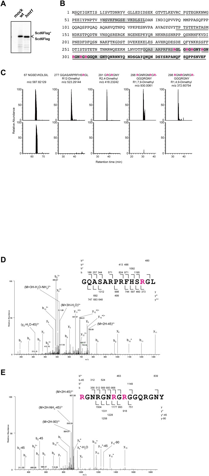
Fig 2. Arginine residues in RGG motifs of Scd6 are dimethylated in a Hmt1-dependent manner.
(A) Scd6Flag proteins from wild-type and hmt1 cells were immunoprecipitated using an anti-Flag antibody and were separated using NU-PAGE gels, and were then visualized by immunoblotting with an anti-Flag antibody. (B) Amino acid sequences of Scd6 are shown and chymotriptic peptides that were identified using Tandem mass spectrometry analyses are indicated by underlines. Red letters indicate asymmetric dimethylarginines and residues of RGG motifs are shown in bold. (C) Chromatograms of four chymotryptic peptides of Scd6 containing asymmetric dimethylarginines were compared between wild-type (upper panel) and hmt1 cells (lower panel). Signal intensities of peptides were normalized to total ion current chromatograms and ratios of signal intensities of peptides between wild-type and hmt1 cells are presented as relative abundances of each peptide. NGSEVKDLSIL was used as an unmodified Scd6 peptide. (D, E) MS/MS spectra of identified peptides containing asymmetric dimethylarginines; GQASARPRFHSRGL (D), RGNRGNRGRGGQRGNY (E).

