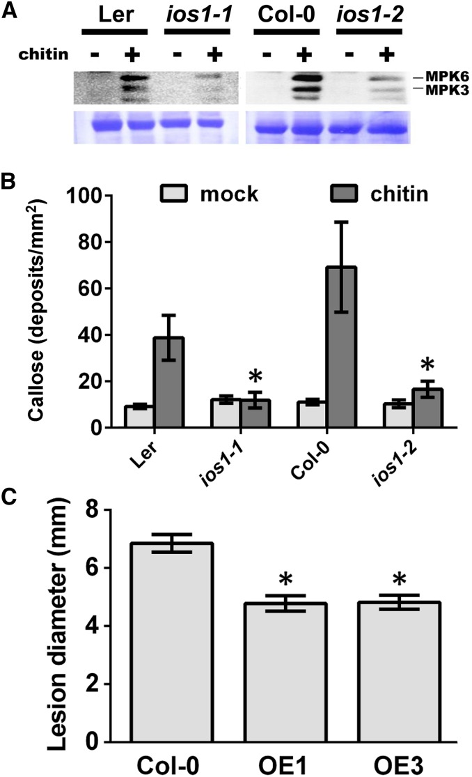
Figure 8.
A Role for IOS1 in the Chitin Response.
(A) MPK activation upon elicitation with chitin. Fourteen-day-old seedlings from Ler-0 or Col-0 wild type, ios1-1, or ios1-2 were syringe-infiltrated with 0.2 mg/mL chitin for 5 min. Immunoblot analysis using phospho-p44/42 MPK antibody is shown in top panel. Lines indicate the positions of MPK3 and MPK6. Coomassie blue staining is used to estimate equal loading in each lane (bottom panel). An independent experiment showed similar results.
(B) Callose deposition upon elicitation with chitin. Fourteen-day-old seedlings from Ler-0 and ios1-1 or Col-0 and ios1-2 were treated with 0.2 mg/mL chitin and samples were collected 16 h later for aniline blue staining. Numbers are averages ± se of callose deposits per square millimeters from two independent experiments each including six seedlings (n = 12). Asterisks indicate a significant difference to wild-type controls based on a paired two-tailed t test (P < 0.01).
(C) B. cinerea-mediated lesions. Arabidopsis leaves of Col-0 and IOS1 overexpression lines were droplet-inoculated (10 μL) with 105 B. cinerea spores/mL and lesion diameters were evaluated at 3 dpi. Data are average ± se of lesion diameters from two independent experiments each with six plants (n = 12). Asterisks indicate a significant difference to wild-type controls based on a paired two-tailed t test (P < 0.01).

