
FIGURE 3.
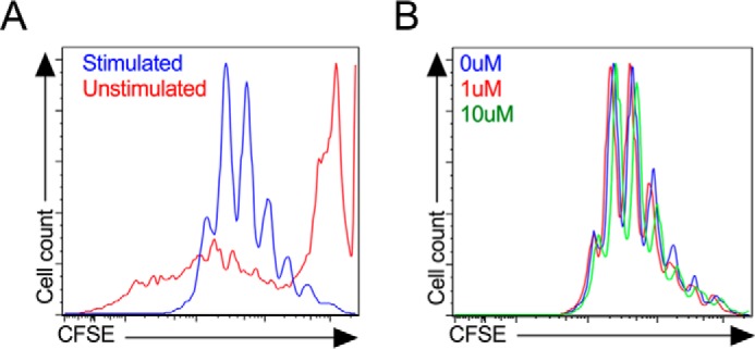
Proliferation of splenocytes in response to pioglitazone treatment in vitro. Unfractionated NOD mouse splenocytes were stimulated in vitro with anti-CD3/anti-CD28 and IL-2 for 4 days then gated on CD4+ cells by flow cytometry. A, CFSE dye fluorescence intensity in unstimulated (red line) and stimulated (blue line) CD4+ cells in the absence of pioglitazone. B, effects of 0 μm (blue line), 1 μm (red line), and 10 μm (green line) pioglitazone on CFSE fluorescence intensity in stimulated CD4+ cells.