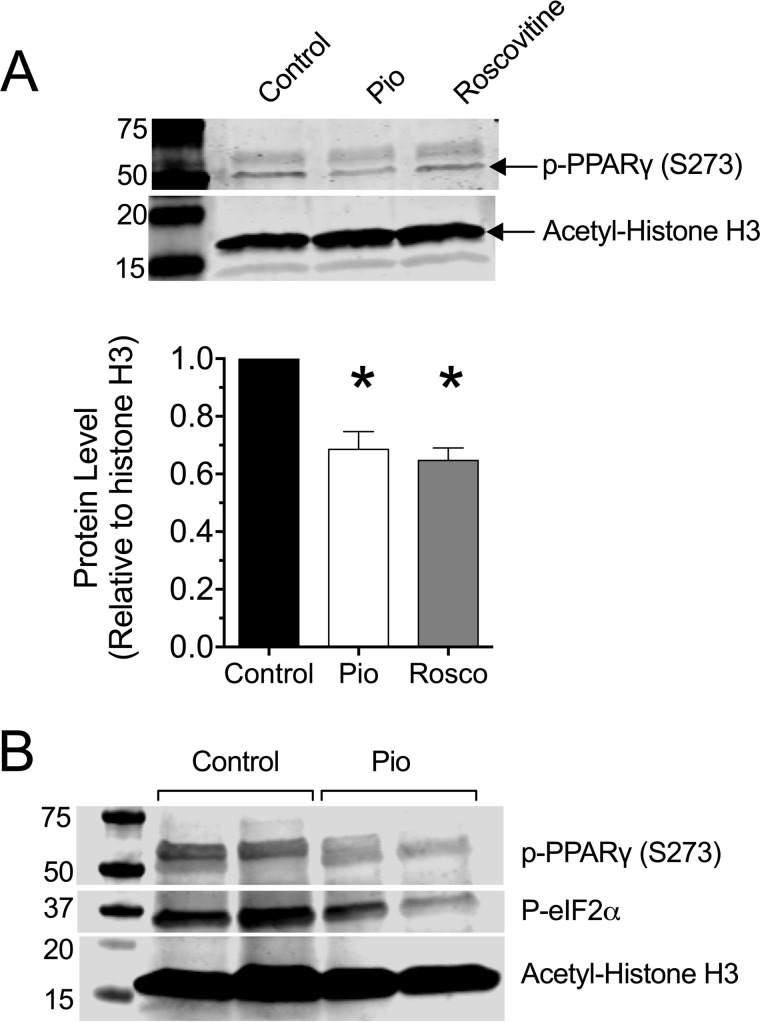
FIGURE 4.
Pioglitazone suppresses PPAR-γ (Ser-273) phosphorylation in β cells in vitro and in islets of NOD mice in vivo. A, MIN6 β cells were preincubated in vehicle, 10 μm pioglitazone (Pio), or 10 μm roscovitine overnight, then extracts were subjected to immunoblotting using anti-phospho-PPAR-γ (S273) and anti-acetyl-histone H3 (Lys-14) as a loading control. Representative immunoblots are shown, and the bar graph below shows the quantitation of immunoblots (normalized to loading control) from three independent experiments. * indicates that the value is significantly different (p < 0.05) compared with vehicle-treated (control) cells. B, 6-week-old pre-diabetic NOD mice were placed on either normal chow (Control) or chow containing 0.01 wt% pioglitazone (Pio). After 4 of weeks feeding, islets from 2 mice per group were isolated and subjected to immunoblotting using anti-phospho-PPAR-γ (S273), anti-phospho-eIF2α, and anti-acetyl-histone H3 (Lys-14) as a loading control.

