FIGURE 1.
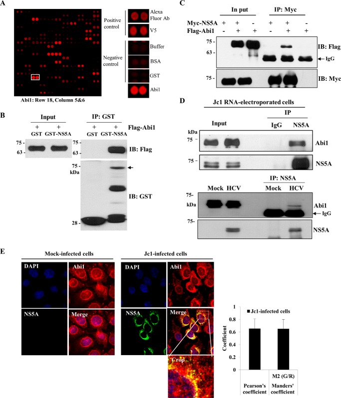
Abi1 is a novel cellular factor interacting with HCV NS5A protein. A, identification of Abi1 in a protein microarray. Both positive and negative controls are shown. B, Abi1 interacts with HCV NS5A protein. HEK293T cells were transiently transfected with FLAG-tagged Abi1 expression plasmid. Total cell lysates harvested at 48 h after transfection were incubated with either GST or GST-NS5A protein. Bound proteins were precipitated with glutathione-Sepharose beads and detected by immunoblotting with an anti-FLAG monoclonal antibody. Protein expressions of GST and GST-NS5A fusion protein were verified by immunoblot analysis using an anti-GST antibody. Arrow indicates GST-NS5A. Input corresponds to 10% of total protein. C, HEK293T cells were transiently transfected with either Myc-tagged NS5A or FLAG-tagged Abi1 expression plasmid, or cotransfected with both plasmids. At 48 h after transfection, cell lysates were immunoprecipitated with an anti-Myc monoclonal antibody, and bound proteins were detected by immunoblot analysis using an anti-FLAG monoclonal antibody (upper panel). Immunoprecipitation efficiency was verified by immunoblot analysis using an anti-Myc antibody (lower panel). D, upper panel, Huh7.5 cells were electroporated with 10 μg of Jc1 RNA and incubated for 3 days. Cell lysates were immunoprecipitated with either IgG or an anti-NS5A antibody and then bound protein was detected by immunoblotting with an anti-Abi1 antibody. Immunoprecipitation efficiency was verified by immunoblot analysis using NS5A antibody. Lower panel, Huh7.5 cells were either mock-infected or infected with Jc1. Total cell lysates were immunoprecipitated with an anti-NS5A antibody. Bound proteins were immunoblotted with an anti-Abi1 antibody. Immunoprecipitation efficiency was verified by immunoblotting with an anti-NS5A antibody. E, Huh7.5 cells were either mock-infected or infected with Jc1 for 4 h. At 3 days postinfection, cells were fixed in cold methanol at −20 °C for 5 min, and immunofluorescence staining was performed using an anti-Abi1 monoclonal antibody and TRITC-conjugated goat anti-mouse IgG to detect Abi1 (red) and a rabbit anti-NS5A antibody and FITC-conjugated goat anti-rabbit IgG to detect NS5A (green). Dual staining showed colocalization of Abi1 and NS5A in the cytoplasm as yellow fluorescence in the merged images. Cells were counterstained with 4′,6-diamidino-2-phenylindole (DAPI) to label nuclei (blue). The enlarged selection marked by a white square is shown as a crop image. Colocalization of Abi1 and HCV NS5A was quantified by both Pearson's and Manders' overlap coefficients. More than 10 cells were applied to ImageJ for quantification of overlap coefficient, and error bars indicate the mean ± S.D. Experiments were performed in duplicate.

