FIGURE 5.
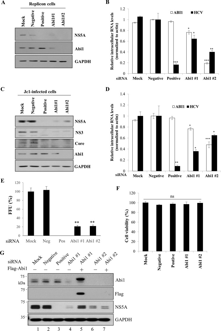
Knockdown of Abi1 suppresses HCV propagation. A, Huh7 cells harboring HCV subgenomic replicon (genotype 1b) were either mock-transfected or transfected with 60 nm of the indicated siRNA constructs. Total cell lysates harvested at 96 h after transfection were immunoblotted with the indicated antibodies. Negative, scrambled siRNA; positive, HCV-specific siRNA; suffixes #1 and #2 refer to the siRNA sequences targeting two different regions of Abi1. B, total RNAs were extracted from siRNA-transfected HCV replicon cells and both intracellular HCV RNA and Abi1 mRNA levels were quantified by qRT-PCR. The asterisks indicate significant differences (*, p < 0.05; **, p < 0.01; ***, p < 0.001) from the value for the negative control. Experiments were carried out in triplicate. Error bars indicate the mean ± S.D. C, Huh7.5 cells were either mock-transfected or transfected with 60 nm of the indicated siRNA constructs. At 48 h after transfection, cells were infected with Jc1 for 4 h. Total cell lysates harvested at 48 h after HCV infection were immunoblotted with the indicated antibodies. D, Huh7.5 cells were either mock-transfected or transfected with the indicated siRNAs and infected with Jc1 at 48 h after transfection. At 96 h after siRNA transfection, both intracellular HCV RNA and Abi1 mRNA levels were quantified by qRT-PCR. The asterisks indicate significant differences (*, p < 0.05; **, p < 0.01; ***, p < 0.001) from the value for the negative control. Experiments were carried out in triplicate. Error bars indicate the mean ± S.D. E, Huh7.5 cells were either mock-transfected or transfected with two different Abi1-specific siRNAs for 48 h and then infected with Jc1 for 4 h. At 2 days postinfection, culture supernatant was harvested and then used to infect naive Huh7.5 cells. HCV infectivity was determined by FFU/ml. Experiments were performed in triplicate. Error bars indicate the mean ± S.D. F, Huh7.5 cells were either mock-transfected or transfected with 60 nm of the indicated siRNAs. At 96 h after transfection, cell viability was determined by WST assay. G, Huh7.5 cells were either mock-transfected or transfected with the indicated siRNAs for 24 h. Cells were further transfected with either empty vector (−) or FLAG-tagged Abi1 plasmid for 24 h and then infected with Jc1. Total cell lysates harvested at 48 h postinfection were immunoblotted with the indicated antibodies. siRNA #1 targets to the 3′ UTR of Abi1, whereas siRNA #2 binds to the coding region of Abi1. Experiments were carried out in triplicate.

