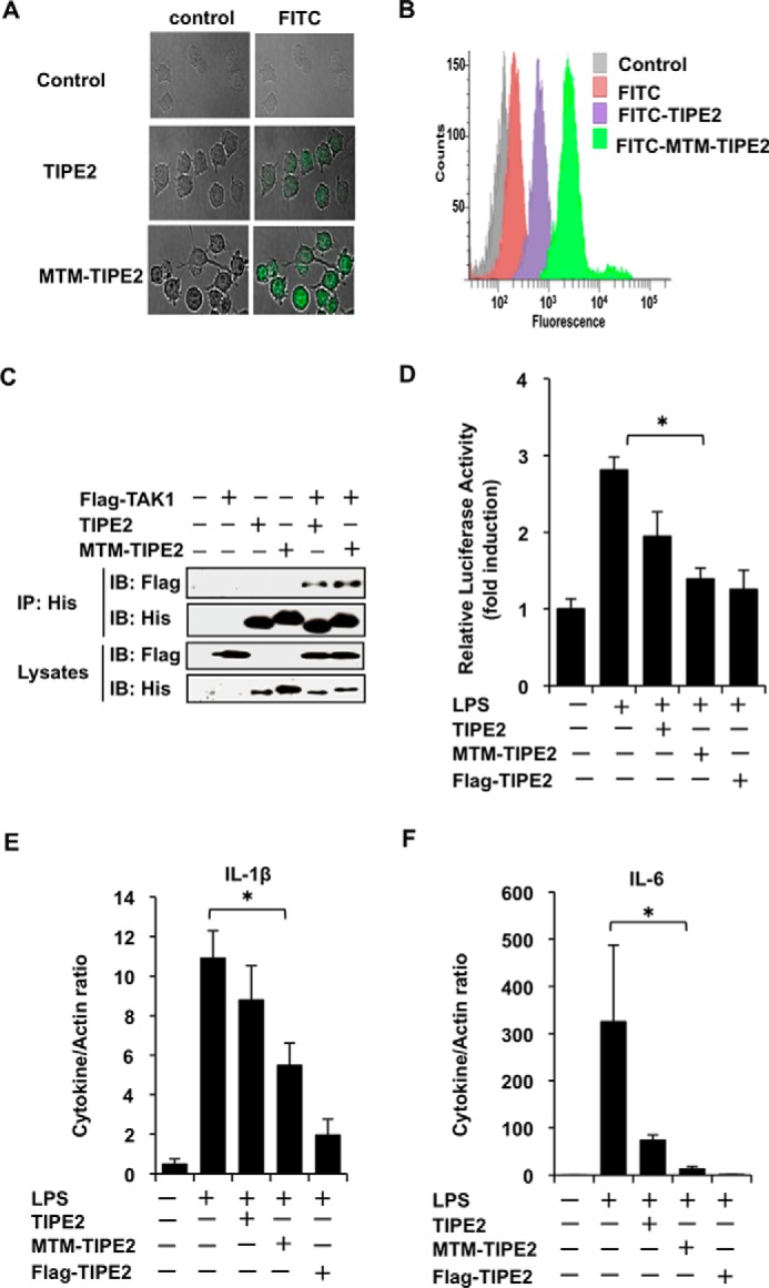
FIGURE 5.

MTM-TIPE2 inhibits LPS-stimulated NF-κB activity and gene expression of inflammatory cytokines in vitro. A and B, RAW246.7 cells were incubated for 30 min in the absence or presence of FITC-labeled MTM-TIPE2 or FITC-labeled TIPE2 at 1 μm, and subsequently the cells were treated for 15 min with proteinase K. Then the cells were observed by confocal microscopy (A) and analyzed by flow cytometry (B). C, the cells were transiently transfected with or without FLAG-TAK1. After 24 h, the cells were incubated for 1 h with or without His-MTM-TIPE2 or His-TIPE2 at 1 μm. The mixtures were used for the His pulldown assay. The bound proteins were detected by Western blotting assay using anti-FLAG and anti-His antibodies. D, cells in 24-well plates were transiently transfected with or without NF-κB reporter construct and then were incubated for an additional 24 h under serum-free culture conditions. Then cells were incubated for 1 h with or without MTM-TIPE2 or TIPE2 at 1 μm. The treated cells were then treated with or without LPS at 100 ng/ml. The cells were lysed after 5 h, and NF-κB reporter activity was measured. FLAG-TIPE2-transfected cells were used as the positive control. The results are expressed as means ± standard deviation in triplicate. *, p < 0.05. E and F, the cells were cultured for 24 h under serum-free culture conditions and then incubated for 1 h with or without MTM-TIPE2 or TIPE2 at 1 μm. Then the cells were treated with LPS at 100 ng/ml. After 2 h, mRNA was prepared, and gene expression of the indicated cytokines was analyzed by qRT-PCR. FLAG-TIPE2-transfected cells were used as the positive control. The results are expressed as means ± standard deviation in triplicate. *, p < 0.05. IB, immunoblot; IP, immunoprecipitation.
