FIGURE 7.
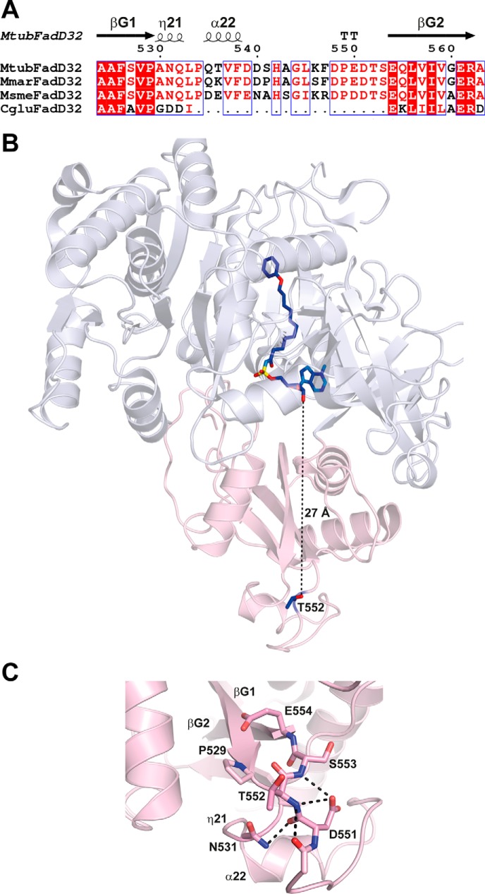
Thr-552 is located in an extended loop fully accessible for phosphorylation by STPKs. A, structure-based sequence alignment of FadD32 from M. tuberculosis, M. marinum, M. smegmatis and Corynebacterium glutamicum in the loop region carrying Thr-552. Sequence homology is highlighted in red, and sequence identity is shown as white letters on a red background. Secondary structure elements (arrows for β-strands and coils for α- and η-helices) are indicated at the top and numbered as in Ref. 35. B, structure of MtbFadD32 (Protein Data Bank code 5HM3). The N- and C-terminal domains are colored blue white and light pink, respectively. The side chain of Thr-552 and the bound PhU-AMS molecule, as found in the structure of MtbFadD32, are shown as a stick representation. C, zoom region of the loop insertion in MtbFadD32 carrying Thr-552. The side chains of residues found within 6 Å of Thr-552 are displayed. Contacts between polar atoms within a distance of 3.5 Å are displayed as dotted lines.

