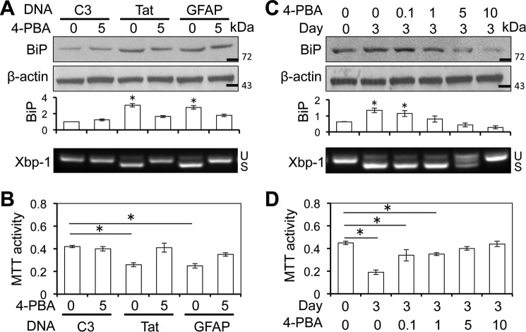
FIGURE 6.
4-PBA inhibited Tat-induced ER stress in astrocytes and astrocyte-mediated Tat neurotoxicity. A and B, U373.MG cells were transfected with cDNA3, Tat, or GFAP expression plasmid; cultured for 48 h; and continued to culture in the presence of 0 and 5 mm 4-PBA for 24 h. A fraction of the cells were harvested and analyzed for BiP expression by Western blotting, and total RNA was isolated from the remaining cells for Xbp-1 alternate splicing by semiquantitative RT-PCR (A). The culture supernatants from the 4-PBA-treated cells were evaluated for neurotoxicity using the MTT assay (B). C and D, iTat primary astrocytes were cultured in the presence of 5 μg/ml Dox for 0 and 3 days and then in the presence of 0, 0.1, 1, 5, or 10 mm 4-PBA for 24 h. A fraction of the cells were harvested and analyzed for BiP expression by Western blotting, and total RNA was isolated from the remaining cells for Xbp-1 alternate splicing by semiquantitative RT-PCR (C). U and S, unspliced and spliced RNA-derived PCR DNA, respectively. The culture supernatants from the 4-PBA-treated cells were evaluated for neurotoxicity using the MTT assay (D). -Fold changes in BiP expression over the control (C3 at day 0) were the mean ± S.D. (error bars) of three independent repeats and are shown below the respective Western blots (A and C). *, p < 0.05.

