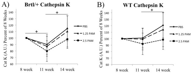
Figure 5.
Near infrared optical imaging of cathepsin K activatable substrate was visualized at 8 weeks. Subsequent imaging of probe administered at 11 and 14 weeks reveals transient reductions (11 wk) and restoration (14 wk) of osteoclast activity, particularly in Brtl/+ mice. Despite large variability, a general dose-dependent trend was noted across doses of pamidronate. + p<0.05 within sample repeated measure ANOVA showing effect of time following cessation.

