Figure 1.
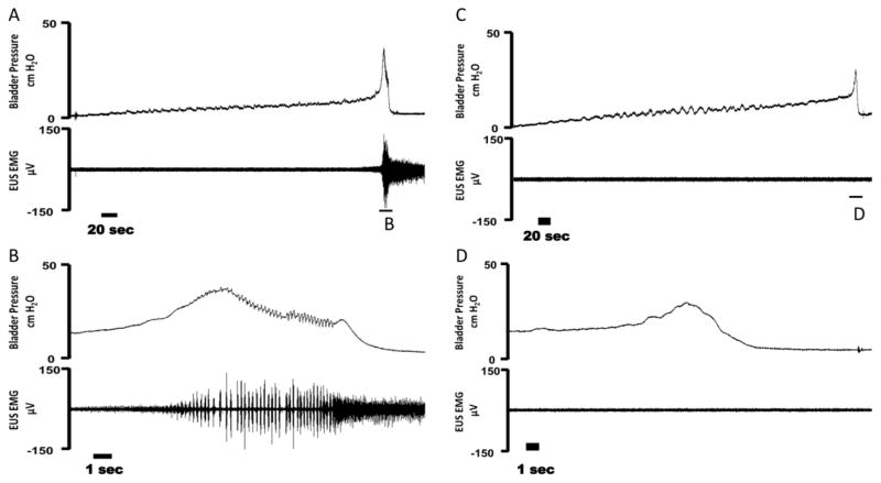
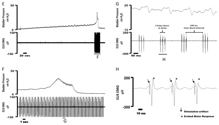
Example cystometrogram (CMG) trials in female rats during intact (A–B), bilateral pudendal motor transection (C–D), and bilateral phasic stimulation of the distal pudendal motor stump (E–H). (A) Bladder pressure (top) and external urethral sphincter (EUS) EMG activity (bottom) during a distention evoked trial. (B) An expanded trace from (A) showing bladder pressure (top) and EUS EMG activity confirming the presence of high frequency oscillations (HFOs) during a void event. (C) Bladder pressure (top) and EUS EMG activity (bottom) during a bilateral pudendal motor transection trial. (D) An expanded trace from (C) showing bladder pressure (top) and EUS EMG activity confirming the absence HFOs during a void event due to motor transection. (E) Bladder pressure (top) and EUS EMG activity (bottom) during bilateral phasic stimulation of the distal pudendal motor stump. Stimulation is turned on during a voiding event and is subsequently turned off after the end of the void. (F) An expanded trace from (E) showing bladder pressure (top) and EUS EMG (bottom) during the bladder contraction. Phasic stimulation of the distal pudendal motor stump elicited HFO’s during stimulation. (G) An expanded trace from (F) of bladder pressure (top) and EUS EMG (bottom) during phasic stimulation. Oscillations in bladder pressure follow each stimulation burst. (H) An expanded trace from (G) of EUS EMG activity during a single phasic burst. Arrow indicates the stimulation artifact and * denotes an evoked motor response.


