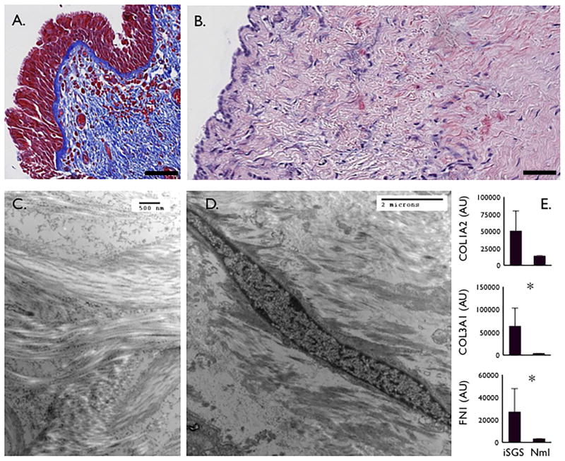
Figure 1. iSGS Anatomic, Physiologic and Histologic characteristics.
Trichome blue staining of iSGS subglottic scar demonstrating extensive blue collagen staining and an intact basement membrane (Magnification 20x, scale bar = 100μM) (A.). Sirius red staining highlights subepithelial collagen deposition in red (Magnification 40x, scale bar = 100μM) (B.). Transmission electron microscopy shows irregular, disordered subepithelial collagen bundles (C.), as well as abundant fibroblasts (D.). qPCR from 10 iSGS patients for extracellular matrix constituents (Types I & III collagen, Fibronectin) from iSGS scar compared with 23 healthy controls (E.), asterisk indicates statistical significance (Two-tailed, Mann Whitney test; p<0.05).

