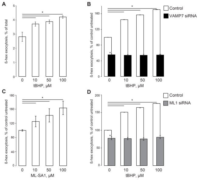
Fig 1. Moderate ROS levels activate lysosomal exocytosis.
A. β-hex exocytosis in cells treated with low and moderate tBHP concentrations. β-hex activity in the extracellular medium expressed as the percentage of total cellular β-hex content at different tBHP concentrations. B. β-hex exocytosis normalized to its levels in untreated cells, which were taken as 100%. VAMP7 knockdown using siRNA suppresses the basal and the tBHP-stimulated β-hex exocytosis. C. β-hex exocytosis expressed as in panel B as a function of ML-SA1 concentration. D. β-hex exocytosis analyzed as in panel B in mock- and TRPML1 siRNA-transfected cells. β-hex assays in HeLa cells performed as in our previous publications [26, 29, 40]. β-hex activity in the culture medium was analyzed one hour after the fresh medium was introduced. The activity was normalized to the total β-hex content, which was analyzed by lysing cells; total β-hex content in cell lysates of untreated cells was taken for 100% (shown in Supplementary Fig S2). tBHP and ML-SA1 were added immediately after the recording had begun. siRNA transfections were performed 24 hours before the experiment. Data represent 3 experiments, 3 independent biological replicates each; * denotes p<0.05 relative to untreated control.

