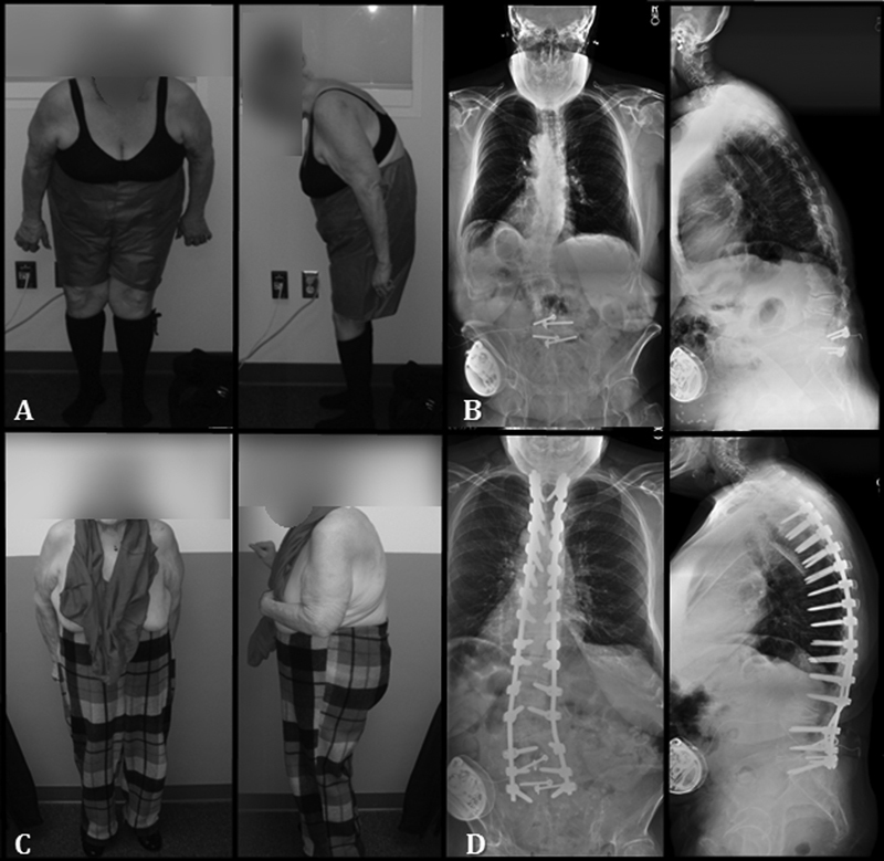
Fig. 1.

(A) Preoperative clinical images and (B) posteroanterior and lateral 36-inch standing radiographs of a 64-year-old woman with a previous L4–S1 fusion who presented with severe lumbar stenosis, neurogenic claudication, and inability to stand upright. (C, D) Post–pedicle subtraction osteotomy clinical and radiographic images showing improvement of both coronal and sagittal alignment.
