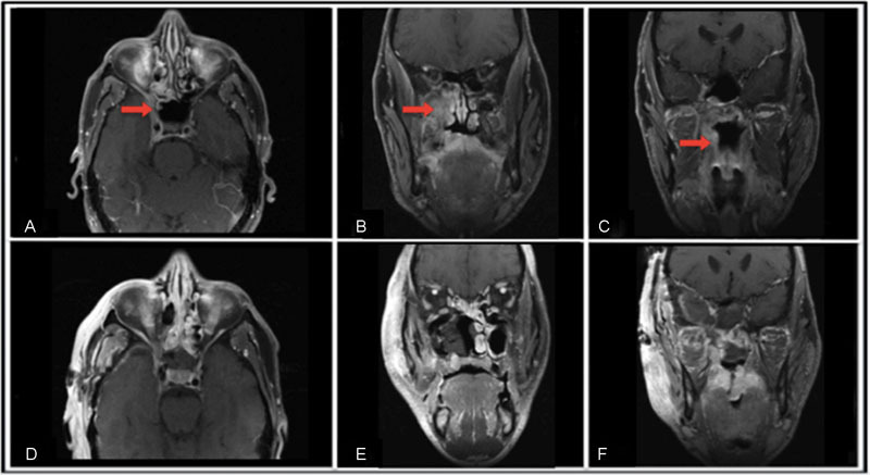
Fig. 3.

Preoperative MRI show a large mass infiltrating the right lateral wall of the nasopharynx, extending to the pterygomaxillary and infratemporal fossa, and reaching the anteromedial aspect of the middle fossa to come in close contact with the temporal pole of the brain: (A) Pre-op axial, (B) Pre-op coronal-A, (C) Pre-op coronal-B. Post-operative MRI shows the resolution of the lesion: (D) Post-op axial, (E) Post-op coronal-A, (F) Post-op coronal-B.
