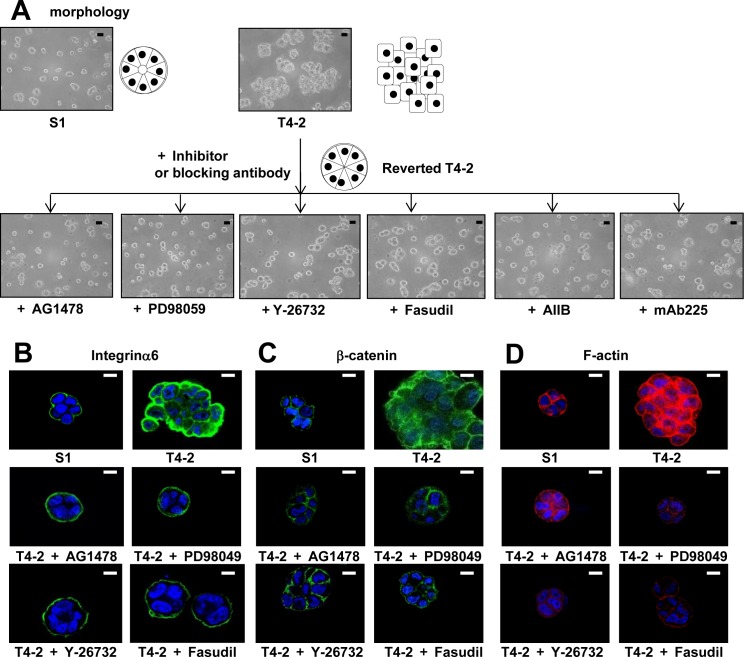
Figure 3. Involvement of ROCK in loss of polarity and actin cytoskeletal rearrangement of T4-2 cells in 3D lrECM.
A. Morphological alternations of T4-2 cells treated with ROCK inhibitor (Y-26732 or Fasudil, 30 μM), EGFR inhibitor (AG1478, 0.1 μM), Integrinβ1 function blocking antibody (AIIB, 100 μg/mL), EGFR function blocking antibody (mAB225, 4 μg/mL) or MEK inhibitor (PD98059, 10 μM). Scale bars: 20 μm. B. C. and D. Confocal immunofluorescence of basal maker, lateral marker and cytoskeletal filamentousF.-actin. Their images were obtained using their specific antibodies (B. Integrinα6; Green, C. β-catenin; Green) and Alexa Fluor-conjugated phalloidin (D.) F-actin; Red). Nuclei were stained with DAP (Blue). Scale bars: 10 μm.

