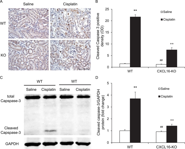
Figure 4. CXCL16 deficiency inhibits Caspase-3 activation in tubular epithelial cells.
A. Representative photomicrographs of kidney sections stained for cleaved caspase-3 (brown) and counterstained with hematoxylin (blue) in WT and CXCL16 KO mice at 72 hours after cisplatin or saline treatment. Scale bar: 50μm. B. Quantitative analysis of cleaved Caspase-3 expression in kidneys of WT and CXCL16 KO mice after cisplatin or saline treatment. **P < 0.01 vs. WT saline; ##P < 0.01 vs. CXCL16 KO cisplatin; ++P < 0.01 vs. WT cisplatin. n = 6 in each group. C. Representative western blots show cleaved Caspase-3 protein expression in kidneys of WT and CXCL16 KO mice after cisplatin or saline treatment. D. Quantitative analysis of cleaved Caspase-3 protein expression in kidneys of WT and CXCL16 KO mice after cisplatin or saline treatment. **P < 0.01 vs. WT saline; #P < 0.05 vs. CXCL16 KO cisplatin; +P < 0.05 vs. WT cisplatin. n = 6 in each group. HPF, high power field; GAPDH, glyceraldehyde-3-phosphate dehydrogenase.

