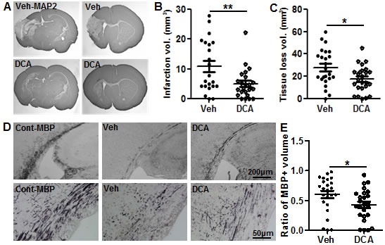
Figure 1. DCA treatment reduced brain injury after HI.

A. Representative MAP2 staining from the dorsal hippocampus (left panels) and striatum (right panels) at 72 h post-HI in vehicle-treated (upper panels) and DCA-treated mice (lower panels). B. The infarction volume at 72 h after HI in DCA-treated (n = 25) and vehicle-treated mice (n = 24). C. The total tissue loss volume at 72 h after HI in DCA-treated and vehicle-treated mice. D. Representative MBP staining at the hippocampal level shows the myelin structure in the subcortical white matter of the ipsilateral hemisphere at 72 h after HI in vehicle-treated and DCA-treated mice as well as in normal control mice. The lower panel in E shows higher magnification of MBP-stained subcortical white matter. E. Quantitative analysis showed the tissue loss in the subcortical white matter in DCA-treated (n = 25) and vehicle-treated mice (n = 24). *p < 0.05, **p < 0.01.
