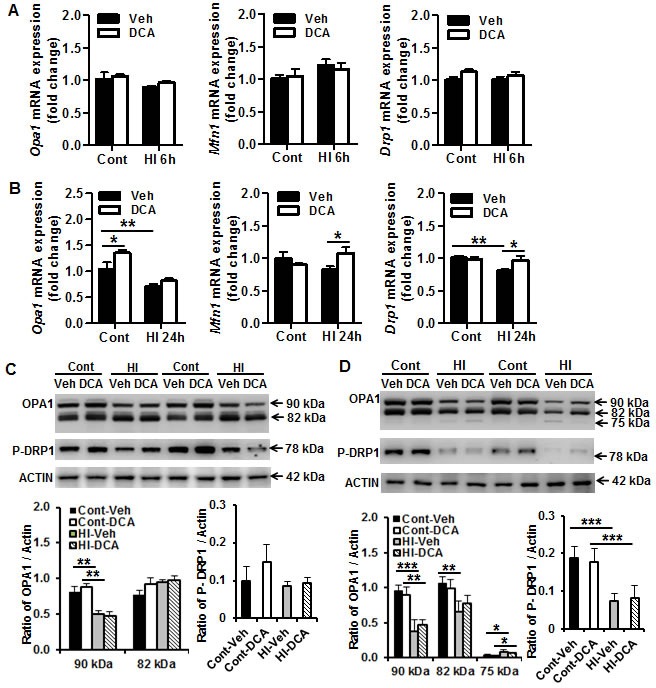
Figure 4. Effect of DCA treatment on brain mitochondrial fission and fusion.

A. The mRNA expression levels of the mitochondrial fusion (Mfn1, OPA1) and fission (Drp1) genes in the mouse brain were quantified by RT-qPCR at 6 h after HI. B. The mRNA expression levels of the mitochondrial fusion (Mfn1, OPA1) and fission (Drp1) genes in the mouse brain were quantified by RT-qPCR at 24 h after HI. n = 5 for the vehicle control, n = 6 for the DCA control at 6 h, n = 6 for the 6 h HI and 6 h DCA groups; n = 6 for the vehicle control, n = 7 for the DCA control at 24 h, n = 8 for the 24 h HI vehicle group, and n = 7 for the 24 h HI DCA group. C. Immunoblotting of OPA1, MFN1, and P-DRP1 in the mitochondrial fraction of normal control (Cont) and 6 h after HI treated with vehicle (Veh) or DCA. The 90 kDa upper band of OPA1 was decreased significantly at 6 h after HI, and DCA treatment did not prevent the reduction (n = 6/group). D. Immunobloting of OPA1, and P-DRP1 in normal controls and 24 h after HI treated with vehicle or DCA. The expression of these proteins decreased significantly at 24 h after HI in the mitochondrial fraction. The 75 kDa cleavage band of OPA1 increased significantly after HI, and DCA treatment has no significant effect on OPA1 cleavage at 24 h after HI (n = 6/group). *p < 0.05, **p < 0.01.
